
Hugh Turvey: Inside the Life of an X-Ray Artist
This post was originally published in January 2014. We’re resurfacing it as part of our #ThrowbackThursday effort to give some love to our favorite posts.—The Proof Team
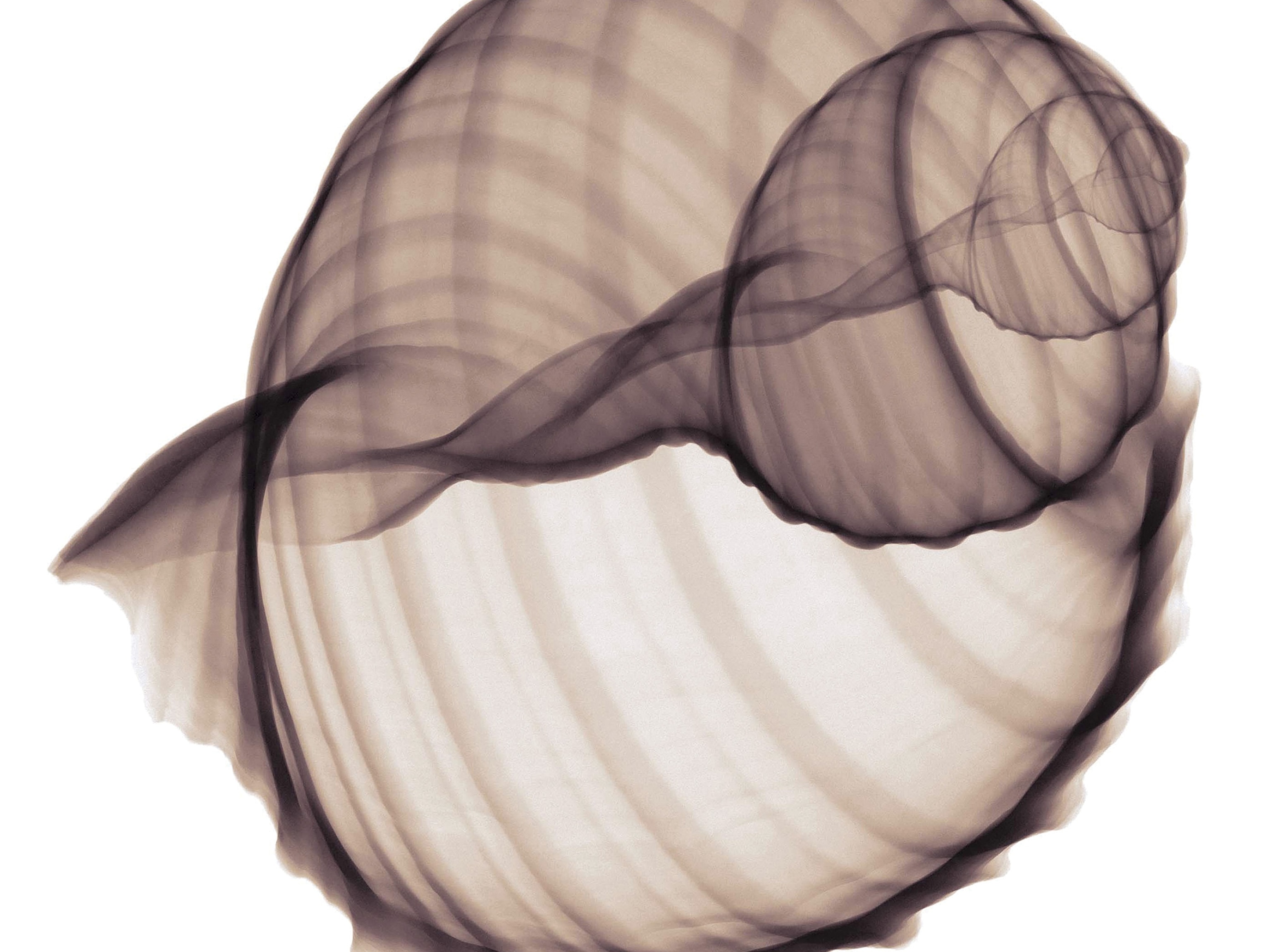
Hugh Turvey is a British artist and photographer who uses x-ray technology to create what he calls xograms, a fusion of visible light and x-ray imagery. I first worked with him for an assignment in the magazine and have to come to rely on his expertise when it comes to seeing the unseen. I spoke with him about his work and recent photo published in the February issue of National Geographic magazine.

“I am an experimentalist and I think in images. Since I started working with x-ray in the late 1990s, I am constantly amazed with how little I know.”—Hugh Turvey
JEANNE MODDERMAN: Tell me about your initial foray into x-ray imaging.
HUGH TURVEY: My x-ray passion was ignited after a designer friend asked for a broken bone image for an album cover. At the time, I was doing a four-year apprenticeship with iconic music photographer Gered Mankowitz in London. I took a walk from his studio in Belsize Park up to the local hospital to meet the head of radiography to seek his advice. (Oh, and yes, I was planning to be a rock-and-roll photographer.)

JEANNE: What is the most challenging aspect of your work?
HUGH: Density defines my work. Very big or very small objects challenge the technology and physics. Commercially we have been asked to x-ray a huge range of subjects over the years. We once had a call from Saatchi Advertising in London wanting to know if I could x-ray a building. In my mind nothing is impossible, but a line has to be drawn somewhere.
The trickiest image was trying to get a series of simultaneous rotating x-ray and visible light objects for an educational eBook and app called X Is for X-Ray. This had never been done before and the technology didn’t really exist. So with a lot of patience and melding of techniques we were able to introduce kids to science in an easily digestible format. I am very proud of this and am continuing work on it.

JEANNE: Do you have a favorite image?
HUGH: One of my personal favorite images is titled “Femme Fatale.” It is a colored x-ray image of a woman’s foot in a stiletto shoe. It is one of my first images—transparent, self-explanatory, and [it] has become an iconic image inadvertently, possibly made more unique by radiation law amendments. It also happens to be my wife and a fitting portrait of my sole mate … pun intended.

JEANNE: Your work has been published a few times in National Geographic magazine. I recently assigned you to x-ray an elephant skull for the February issue. Can you talk about the process?
HUGH: Have you ever tried to get an elephant skull? Have you ever tried to lift an elephant skull? It turns out that I have now, and needless to say it was not easy.
We are accustomed to sourcing specimens and had recently imaged a huge python for National Geographic and a series of peregrine falcons for Discovery. However, an elephant skull with good teeth in the mandible was a little more elusive. We were put in contact with a company who works to ensure the welfare of nondomesticated, wild animals which have been abandoned, confiscated, or surrendered. They had not only a skull and mandible, but also the tusks, a collection of ivory and skin items, and an elephant foot table. (The total insurance value of these objects was nearly $150,000!) To lift the skull required three people, and two for the mandible.
Normal medical equipment is very finely calibrated to the human body and useless for this application. We were forced to upgrade to industrial x-ray, which is normally used for x-raying engineering structures, such as bridges.

JEANNE: Can you talk about your post-process work? What goes into making the finished product?
HUGH: We have huge pieces of film that need digitizing using photographic scanners. These are layered and combined to produce a grayscale image at approximately one gigabite. Coloring is where you can interject depth back into the image and control the path of the eye over the image. Even though I have written the smallest reply to this question, post-process accounts for at least 70 percent of my time!

Hugh Turvey’s solo show “X-POSÉ: Material and Surface” will be on display at the Oxo Tower Wharf in London from February 12 to 23. Follow Jeanne Modderman on Twitter and Instagram.
Related Topics
You May Also Like


Go Further
Animals
- How can we protect grizzlies from their biggest threat—trains?How can we protect grizzlies from their biggest threat—trains?
- This ‘saber-toothed’ salmon wasn’t quite what we thoughtThis ‘saber-toothed’ salmon wasn’t quite what we thought
- Why this rhino-zebra friendship makes perfect senseWhy this rhino-zebra friendship makes perfect sense
- When did bioluminescence evolve? It’s older than we thought.When did bioluminescence evolve? It’s older than we thought.
- Soy, skim … spider. Are any of these technically milk?Soy, skim … spider. Are any of these technically milk?
Environment
- Are the Great Lakes the key to solving America’s emissions conundrum?Are the Great Lakes the key to solving America’s emissions conundrum?
- The world’s historic sites face climate change. Can Petra lead the way?The world’s historic sites face climate change. Can Petra lead the way?
- This pristine piece of the Amazon shows nature’s resilienceThis pristine piece of the Amazon shows nature’s resilience
- Listen to 30 years of climate change transformed into haunting musicListen to 30 years of climate change transformed into haunting music
History & Culture
- Meet the original members of the tortured poets departmentMeet the original members of the tortured poets department
- Séances at the White House? Why these first ladies turned to the occultSéances at the White House? Why these first ladies turned to the occult
- Gambling is everywhere now. When is that a problem?Gambling is everywhere now. When is that a problem?
- Beauty is pain—at least it was in 17th-century SpainBeauty is pain—at least it was in 17th-century Spain
Science
- Here's how astronomers found one of the rarest phenomenons in spaceHere's how astronomers found one of the rarest phenomenons in space
- Not an extrovert or introvert? There’s a word for that.Not an extrovert or introvert? There’s a word for that.
- NASA has a plan to clean up space junk—but is going green enough?NASA has a plan to clean up space junk—but is going green enough?
- Soy, skim … spider. Are any of these technically milk?Soy, skim … spider. Are any of these technically milk?
Travel
- Dina Macki on Omani cuisine and Zanzibari flavoursDina Macki on Omani cuisine and Zanzibari flavours
- How to see Mexico's Baja California beyond the beachesHow to see Mexico's Baja California beyond the beaches
- Could Mexico's Chepe Express be the ultimate slow rail adventure?Could Mexico's Chepe Express be the ultimate slow rail adventure?
